How PCR Powers Modern DNA Sequencing Technology
How PCR Powers Modern DNA Sequencing Technology
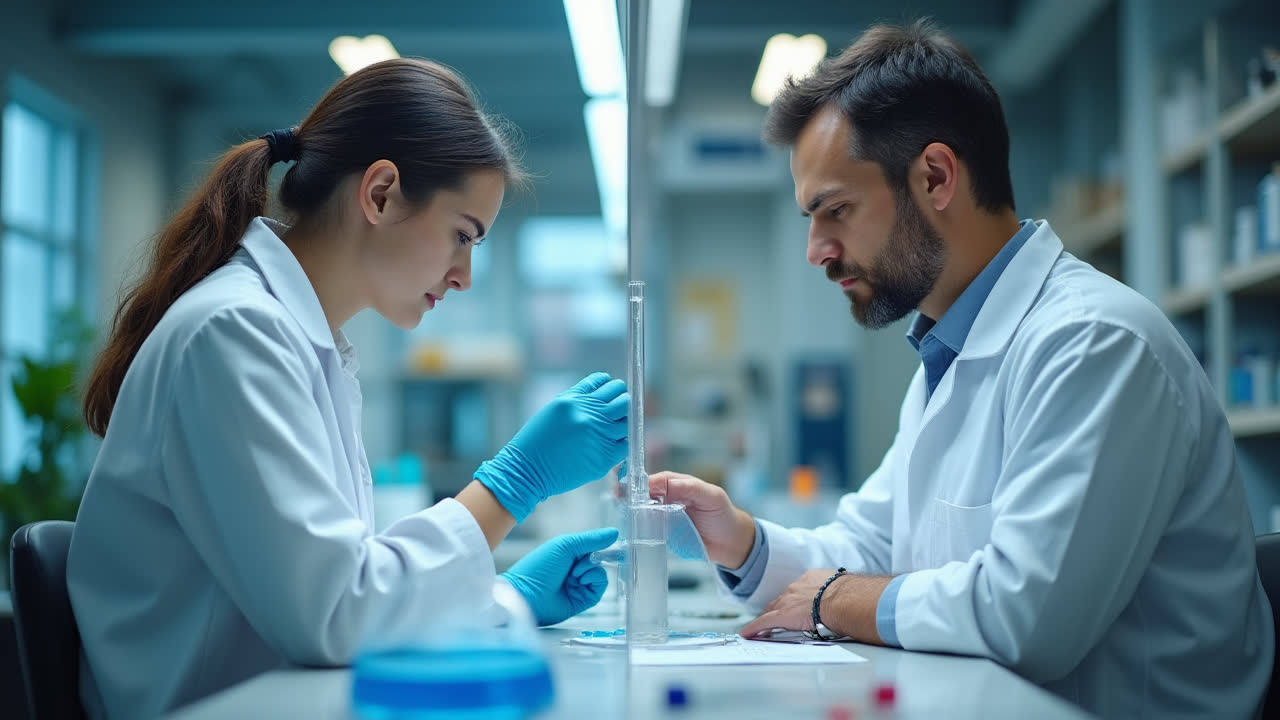
The revolutionary technique of Polymerase Chain Reaction (PCR) plays a pivotal role in DNA sequencing, enabling scientists to decode the genetic blueprint of organisms. Have you ever wondered how scientists determine the precise order of nucleotides in DNA? The answer lies in the remarkable partnership between PCR technology and sophisticated sequencing methods. This article explores the essential relationship between PCR and DNA sequencing techniques, illuminating how this molecular tool has transformed genetic analysis.
Understanding DNA Sequencing Fundamentals
DNA sequencing represents one of the most significant technological breakthroughs in modern molecular biology. At its core, sequencing is a laboratory technique used to determine the precise order of nucleotides (adenine, guanine, cytosine, and thymine) within a DNA molecule. This information serves as the foundation for countless applications in medical research, forensics, evolutionary biology, and personalized medicine.
The journey of DNA sequencing began with Frederick Sanger's groundbreaking method in 1975, which revolutionized our ability to read genetic information. Since then, technological advances have dramatically improved sequencing capabilities, leading to faster, more affordable, and increasingly accurate methods. What remains constant throughout this evolution is the critical role of PCR in preparing and amplifying DNA for sequencing protocols.
Modern DNA sequencing typically follows a methodical process: sample preparation, DNA extraction, fragmentation, PCR amplification, and finally, the actual sequencing reaction followed by data analysis. Each step builds upon the previous one, creating a sophisticated workflow that converts biological material into readable genetic information. The integration of PCR within this process addresses a fundamental challenge in sequencing - having sufficient quantities of identical DNA fragments to analyze.
The Critical Role of PCR in DNA Sequencing
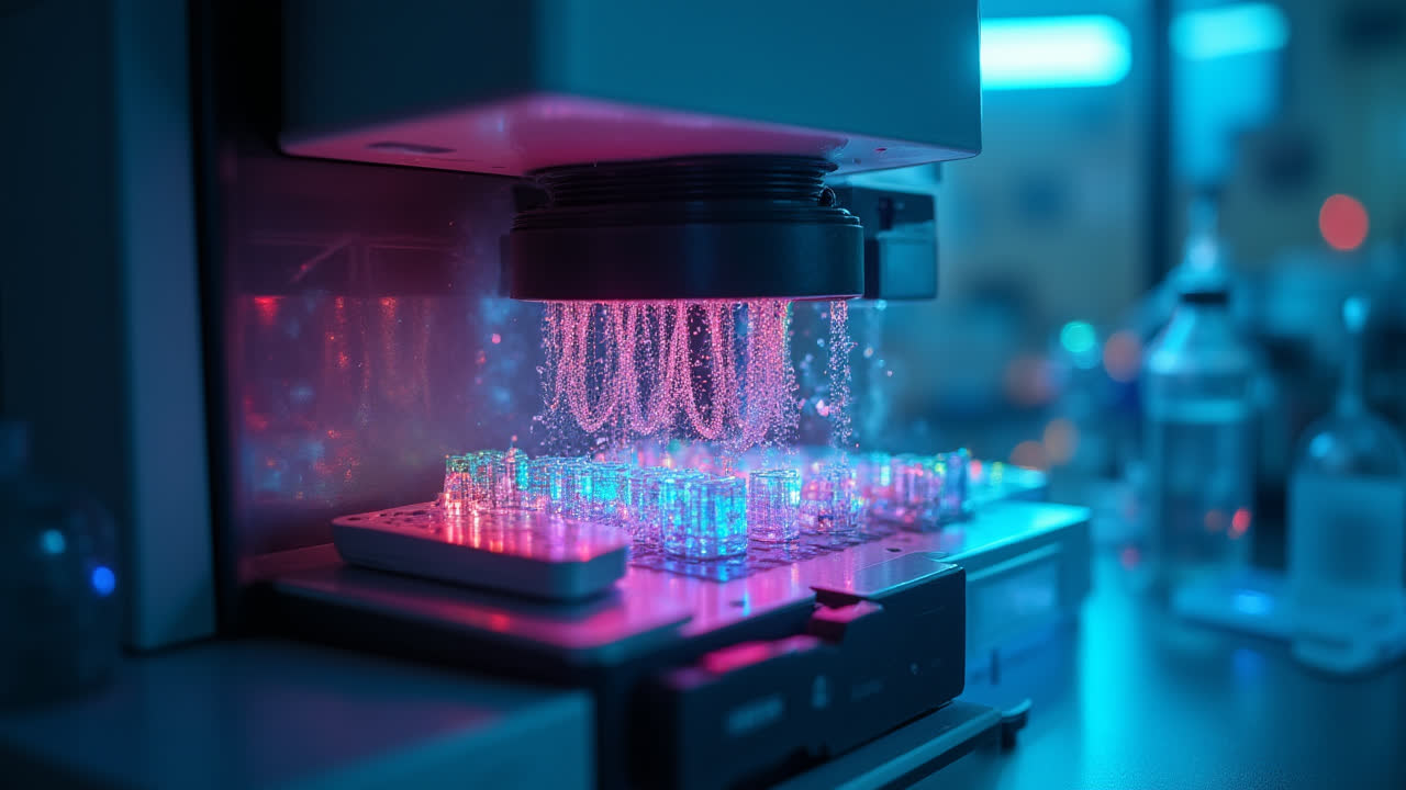
PCR serves as the amplification engine that powers modern DNA sequencing technologies. This ingenious technique, developed by Kary Mullis in the 1980s, creates millions of copies of specific DNA fragments, providing abundant material for sequencing reactions. Without PCR, many sequencing applications would be practically impossible due to the minute quantities of DNA typically available in biological samples.
In sequencing workflows, PCR fulfills several crucial functions. First, it generates the necessary quantity of DNA required for accurate sequencing. Second, it provides the mechanism for incorporating fluorescent markers into DNA fragments - a key feature that enables automated detection systems to identify specific nucleotides. Finally, PCR can be modified to selectively amplify regions of interest, allowing researchers to focus their sequencing efforts on specific genomic sections.
The marriage between PCR and sequencing techniques is particularly evident in the introduction of fluorescently labeled nucleotides. During PCR amplification, modified nucleotides carrying fluorescent tags are incorporated into the growing DNA strands. These fluorescent markers subsequently enable automated sequencing machines to detect and identify each nucleotide as it's incorporated, converting chemical information into digital data that computers can process and analyze.
I recall working in a molecular biology lab where we routinely used PCR to prepare samples for sequencing. The transformation from invisible molecular reactions to colorful fluorescent peaks on a computer screen always seemed like a kind of scientific magic - though it's actually just clever biochemistry at work!
Sanger Sequencing: The Classic Approach
Sanger sequencing, also known as the chain-termination method, represents the first widely adopted DNA sequencing technology. Named after its developer, Frederick Sanger, this method revolutionized genetic analysis and remained the gold standard for decades. Even today, Sanger sequencing maintains relevance for specific applications despite the emergence of newer technologies.
The core principle of Sanger sequencing involves the selective incorporation of modified nucleotides called dideoxynucleotides (ddNTPs) during DNA synthesis. These specialized nucleotides lack the 3'-OH group necessary for forming phosphodiester bonds with subsequent nucleotides. When a ddNTP is incorporated into a growing DNA chain during PCR, extension terminates at that specific position. By controlling the ratio of regular deoxynucleotides (dNTPs) to ddNTPs, researchers generate DNA fragments of varying lengths that terminate at different positions.
In traditional Sanger sequencing, four separate PCR reactions are performed, each containing one type of fluorescently labeled ddNTP (ddATP, ddGTP, ddCTP, or ddTTP). The resulting fragments are separated by size using gel electrophoresis, and the sequence is determined by reading the fluorescent signals in order of increasing fragment length. Modern automated Sanger sequencing typically combines all four ddNTPs in a single reaction, with each type labeled with a different fluorescent dye for simultaneous detection.
Sanger sequencing remains valuable for validating genetic variants, sequencing short DNA regions, and analyzing difficult templates that newer technologies struggle with. Its reliability and accuracy for sequences up to about 1,000 base pairs ensure its continued use in many research and clinical laboratories, particularly when verifying results obtained from other methods.
Next-Generation Sequencing: PCR at Massive Scale
Next-Generation Sequencing (NGS) represents a quantum leap in DNA sequencing capabilities, enabling researchers to analyze millions or even billions of DNA fragments simultaneously. This massive parallelization has dramatically reduced both the time and cost of sequencing entire genomes, making large-scale genetic studies feasible. Despite the technological advances, PCR remains fundamental to most NGS workflows.
In NGS platforms, PCR serves several essential functions. During library preparation, PCR amplifies DNA fragments attached to specialized adapters, creating clusters of identical molecules that function as reaction sites for sequencing. This cluster generation process, sometimes called bridge amplification, produces localized colonies of identical DNA fragments that emit strong enough fluorescent signals for detection when sequencing reactions occur.
Unlike Sanger sequencing with its four separate reactions, NGS methods often employ a sequencing-by-synthesis approach where all four nucleotides, each carrying different fluorescent labels, compete for incorporation during each cycle. The sequence is determined by detecting which fluorescent signal appears at each position as the DNA strand extends. This process occurs simultaneously across millions of clusters, generating massive amounts of sequence data in parallel.
One key difference between Sanger sequencing and NGS is the separation method. While Sanger sequencing typically relies on gel electrophoresis, NGS platforms often employ more sophisticated approaches like capillary electrophoresis or direct imaging of fluorescent signals from immobilized DNA colonies. These methods allow for higher throughput and automation, further increasing sequencing efficiency.
Fluorescent Markers: The Bridge Between PCR and Sequencing
Fluorescent markers represent the critical connection between PCR amplification and sequence detection. These molecular tags convert nucleotide incorporation events into detectable light signals that sequencing instruments can record and interpret. The integration of fluorescent chemistry with DNA replication fundamentally enables modern automated sequencing technologies.
During sequencing reactions, four types of fluorescently labeled ddNTPs are typically used, each carrying a distinct fluorophore that emits light at a specific wavelength. For example, ddATP might be labeled with a green fluorescent dye, ddGTP with yellow, ddCTP with blue, and ddTTP with red. When these modified nucleotides are incorporated into growing DNA strands during PCR, they terminate chain extension and simultaneously mark the position with their characteristic fluorescent signature.
The incorporation of these fluorescent ddNTPs occurs during the PCR process, as DNA polymerase enzymatically adds nucleotides to extend primer-template duplexes. The competition between regular dNTPs (which allow continued extension) and fluorescent ddNTPs (which terminate growth) creates collections of fragments that differ in length by exactly one nucleotide. When separated and detected, these fragments reveal the precise sequence of the original DNA template.
Automated sequencers use laser excitation to cause the fluorescent dyes to emit light, which is then detected by sensitive cameras or photomultiplier tubes. Computer software converts these fluorescent signals into color-coded peaks on an electropherogram, which is then translated into the familiar sequence of A, G, C, and T bases that comprise the DNA sequence. This elegant detection system has enabled the automation and scaling of sequencing technologies that drive modern genomics.
Comparison: Sanger Sequencing vs. Next-Generation Sequencing
| Feature | Sanger Sequencing | Next-Generation Sequencing |
|---|---|---|
| PCR Approach | Four separate reactions with different ddNTPs | Massively parallel PCR with all nucleotides present |
| Throughput | Low (hundreds to thousands of bases) | High (millions to billions of bases) |
| Cost per Base | Higher | Lower |
| Read Length | Longer (700-1000 base pairs) | Shorter (50-600 base pairs typically) |
| Accuracy | Very high (>99.99%) | High but variable (98-99.9% depending on platform) |
| Separation Method | Gel electrophoresis | Capillary electrophoresis or direct imaging |
| Equipment Requirements | Relatively simple | Complex and expensive |
| Applications | Targeted gene sequencing, validation | Whole genome sequencing, transcriptomics, metagenomics |
Frequently Asked Questions
Why is PCR necessary for DNA sequencing?
PCR is essential for DNA sequencing because it serves multiple critical functions. First, it amplifies the target DNA sequence, creating millions of copies from a minute sample to provide sufficient material for sequencing reactions. Second, PCR provides the mechanism for incorporating fluorescently labeled nucleotides or primers that enable automated detection of each base in the sequence. Without PCR, most modern sequencing applications would be impossible due to the tiny amounts of DNA typically available in biological samples and the technical requirements of sequencing instruments.
What's the difference between dNTPs and ddNTPs in DNA sequencing?
dNTPs (deoxynucleotide triphosphates) are the standard building blocks of DNA that contain a 3'-OH group allowing continued chain extension during DNA synthesis. In contrast, ddNTPs (dideoxynucleotide triphosphates) lack the 3'-OH group, which prevents the formation of a phosphodiester bond with the next nucleotide. When a ddNTP is incorporated during PCR, it terminates DNA chain growth at that specific position. In sequencing reactions, fluorescently labeled ddNTPs are used to generate fragments that terminate at different positions, allowing researchers to determine the sequence by analyzing fragment lengths. The competition between dNTPs (which allow continued synthesis) and ddNTPs (which cause termination) creates the collection of fragments needed for sequence analysis.
How do fluorescent markers work in DNA sequencing?
Fluorescent markers in DNA sequencing function by attaching distinct fluorescent dyes to different nucleotides (either directly to ddNTPs or to primers). During sequencing reactions, these labeled nucleotides are incorporated into DNA strands during PCR amplification. Each type of nucleotide (A, G, C, T) is tagged with a different fluorophore that emits light at a specific wavelength when excited by a laser. As DNA fragments pass through a detection system, the sequencing machine records the fluorescent signals in order, generating a chromatogram where each colored peak represents a specific nucleotide. This elegant system enables automated sequence determination without requiring separate reactions for each nucleotide type, dramatically increasing efficiency and throughput compared to earlier methods.
Conclusion
The integration of PCR technology with DNA sequencing methods has fundamentally transformed our ability to decode genetic information. From Sanger's breakthrough chain-termination method to today's high-throughput next-generation platforms, PCR has remained the essential amplification engine that makes sequencing possible. The critical role of PCR in incorporating fluorescent markers during DNA synthesis enables the automated detection systems that have revolutionized genomic research.
As sequencing technologies continue to evolve, with emerging third-generation methods promising even longer read lengths and reduced amplification bias, the relationship between PCR and sequencing may change. However, the fundamental principles established through the PCR-sequencing partnership will likely influence genetic analysis for years to come. This remarkable synergy between amplification and detection techniques exemplifies how combining different molecular methods can lead to transformative scientific capabilities.
Understanding the vital connection between PCR and DNA sequencing not only illuminates the technical foundations of modern genomics but also highlights the creative problem-solving that drives scientific advancement. The journey from basic PCR reactions to decoding entire genomes represents one of molecular biology's most important success stories - one that continues to unfold as researchers develop new methods to read the book of life with increasing speed, accuracy, and affordability.